
Product overview
Sinus Pathways

Sinus Pathways is an anatomical teaching aid for examining the structures and features described below.
Product features and anatomical detail
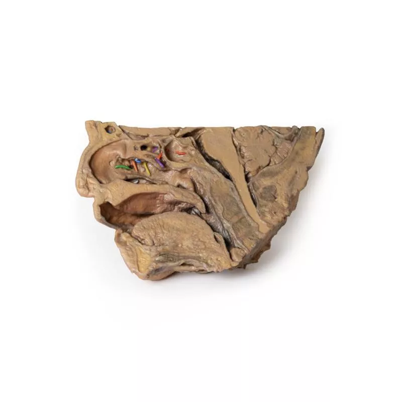
- This 3D model shows a midsagittal to parasagittal section of the right head, highlighting the drainage pathways of the paranasal sinuses into the nasal cavity using colored markers.
- Anteriorly, the nasolacrimal duct (white) opens beneath the inferior nasal concha.
- The middle concha has been sectioned to reveal the maxillary sinus opening via the semilunar hiatus (green), and the drainage of the frontal sinus (blue), anterior (orange) and middle (yellow) ethmoidal cells.
- Posterior ethmoidal cells open into the superior meatus (purple), while the sphenoid sinus drains above the nasopharynx (red).
- The model also preserves key anatomical structures.
- The nasal cavity from nostril to auditory tube, the soft palate, uvula, and pharynx down to the epiglottis.
- The oral cavity is sectioned to show genioglossus and geniohyoid muscles.